ACCEPTED POSITION PAPER · EXPLAINS 2026
Localization Gains from Explanation Regularization Should Not Be Interpreted as Faithfulness Gains
Accepted at the 3rd International Conference on Explainable AI for Neural and Symbolic Methods.
EQUR-XSM · RESEARCH OUTPUTS
ACCEPTED POSITION PAPER · EXPLAINS 2026
Accepted at the 3rd International Conference on Explainable AI for Neural and Symbolic Methods.
In this brain-tumour MRI case study, Contrastive Grad-CAM Consistency (CGC) yields higher mean localisation scores than a cross-entropy baseline. The perturbation-based faithfulness metrics disagree: insertion favours CGC, deletion favours the baseline, and IROF is unchanged at the reported precision.
Study: 3,064 MRI slices from 233 patients · three tumour classes · single-channel ResNet-50 · five-fold patient-grouped cross-validation · fixed training seed.
| Property | Metric | Baseline | CGC |
|---|---|---|---|
| Predictive performance | Accuracy (%) ↑ | 93.63 ± 1.91 | 94.87 ± 1.56 |
| Localisation | Pointing Game (%) ↑ | 27.24 ± 3.76 | 36.02 ± 4.00 |
| Localisation | Relevance Mass Accuracy (%) ↑ | 11.17 ± 1.60 | 18.01 ± 2.70 |
| Faithfulness | Insertion AUC ↑ | 0.73 ± 0.06 | 0.78 ± 0.07 |
| Faithfulness | Deletion AUC ↓ | 0.36 ± 0.07 | 0.41 ± 0.07 |
| Faithfulness | Iterative Removal of Features ↑ | 0.57 ± 0.04 | 0.57 ± 0.07 |
↑ Higher is preferred; ↓ lower is preferred. Comparisons are descriptive: no statistical significance claim is made.
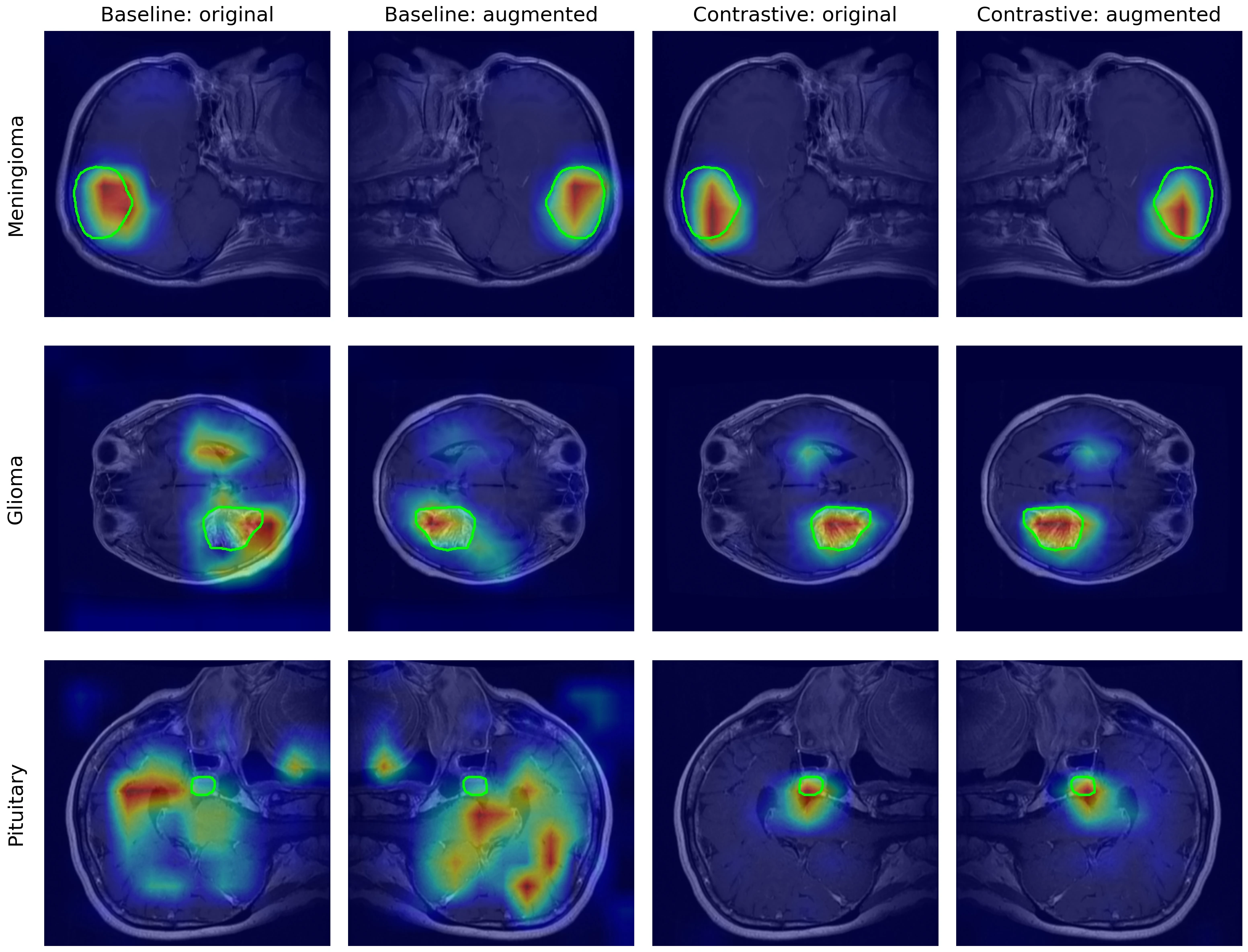
Report transformation consistency, localisation and model reliance as separate evaluation targets. Improvement in one does not establish improvement in the others.
Scope: one 2-D MRI dataset, one backbone, one attribution method and one training seed. Transformation consistency was not measured quantitatively. Faithfulness scores depend on the perturbation protocol; the dataset has documented non-tumour cues. This is a methodological case study, not clinical validation or a new XAI method.
Source: the authors’ accepted camera-ready manuscript, Tables 1–2 and qualitative comparison figure. Supported by ANR-25-CE45-1573 (EQUR-XSM); computations used GRICAD. A public paper link will be added when available.